Cavum septum pellucidum and vergae Image

In the pediatric population, the cavum septum pellucidum is normally seen in all premature infants, in 85% of full-term neonates, and 12% of children between 6 months and 16 years of age. 13. The reported prevalence of persistent cavum septum pellucidum and cavum vergae in adults varies based on the definition used and the modality of diagnosis.. Scatterplot of Cavum Septum Pellucidum and Cavum Vergae (CSPV) Length and Processing Speed (PSS) Score and Psychomotor Speed (PsychoS) Score Among 251 Fighters With CSPV Length Available. eFigure 4. Example of a Patient With Increased Cavum Septum Pellucidum/Cavum Vergae Over Time.
Cavum vergae Ars Neurochirurgica

Cavum septi pellucidi et vergae pacs

Cavum septi pellucidi and cavum vergae MedLink Neurology
Cavum septi pellucidi and cavum vergae MedLink Neurology

Cavum Septum Pellucidum Et Vergae

Cavum Vergae
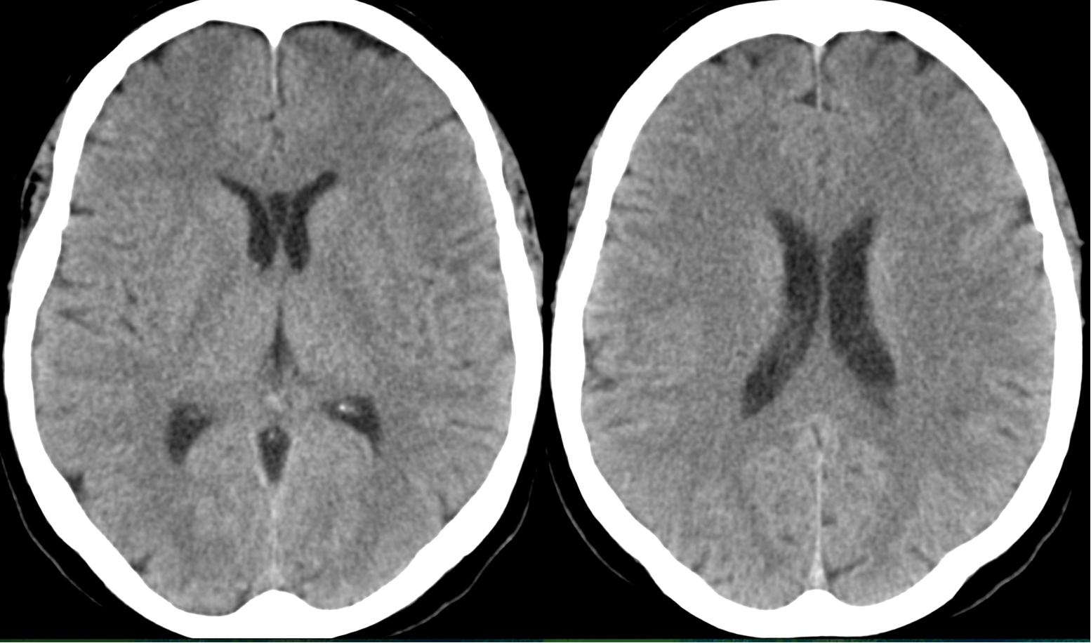
Cavum septum pellucidum, cavum vergae, and cavum veli interpositi (annotated CT) Image
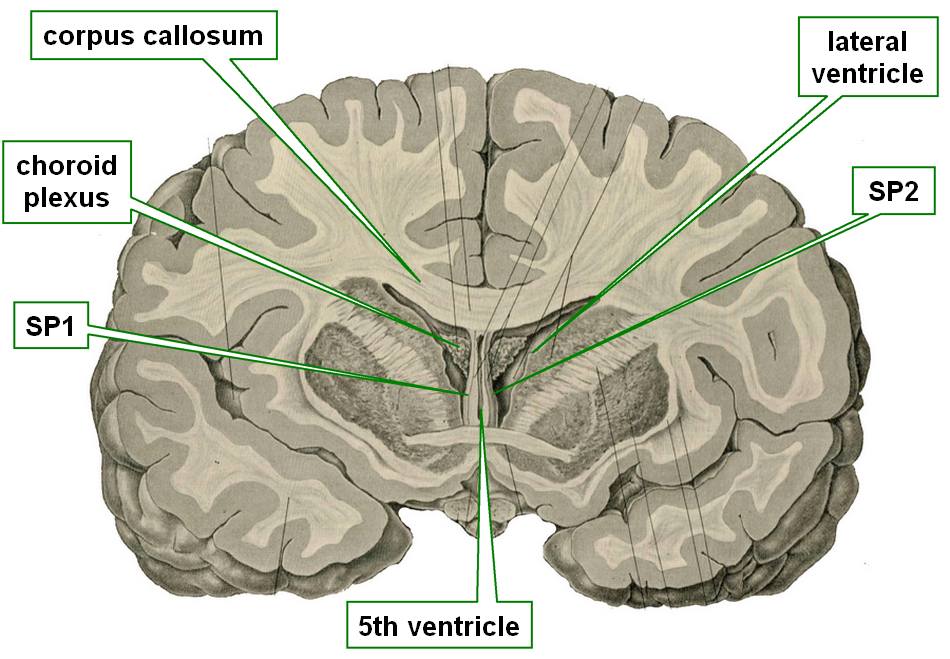
Septum Pellucidum And Fornix

MRI Cavum Septum Pellucidum et Vergae Stock Image C043/0390 Science Photo Library

Cavum Septi Pellucidi and Cavum Vergae With Increased Amyloid β Cortical Load in a 65YearOld

Figure 2 from Cavum septum pellucidum (CSP), Cavum vergae and Cavum veli interpositi Semantic

Cavum Septi Pellucidi (CSP) Brain imaging

Know Your Brain Septum — Neuroscientifically Challenged

Cavum Vergae

Cavum septum pellucidum, cavum vergae, and cavum veli interpositi (annotated CT) Image
.jpg)
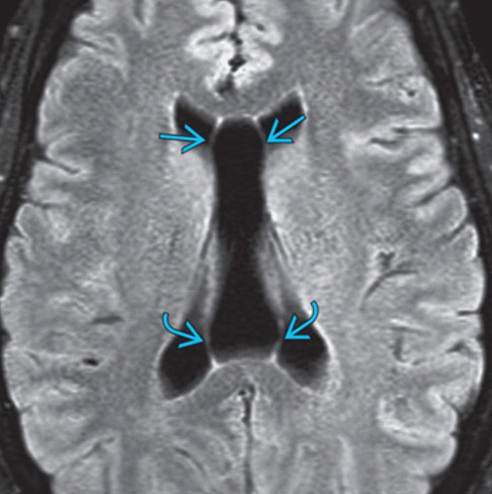
Cavum septum pellucidum and cavum vergae (Radiopaedia 7779790060 Axial Brain Window) NC Commons
Cavum Septi Pellucidi (CSP) Brain imaging

MRI Cavum Septum Pellucidum et Vergae Stock Image C043/0388 Science Photo Library
.jpg)
Cavum septum pellucidum and cavum vergae (Radiopaedia 7779790060 Axial Brain Window) NC Commons
Cavum septum pellucidum and cavum vergae Image
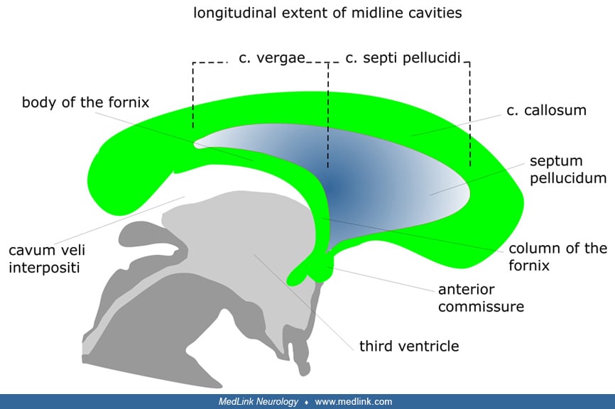
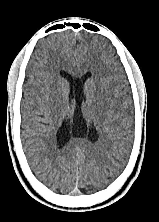
Cavum septum pellucidum (CSP) cyst, cavum vergae, and cavum velum interpositum are various presentations of benign midline anterior intracranial cysts. They are pathological when symptomatic, which arise depending upon the size of the cysts. Cavum septum pellucidum cysts are rare lesions with an incidence of 0.04% . Symptomatic cysts of CSP are.. Cavum Septi Pellucidi, Cavum Vergae, and Cavum Veli Interpositi. The septum pellucidum forms the medial walls of the lateral ventricles and consists of 2 thin laminae, which normally fuse shortly after birth. If the laminae fail to fuse, the potential space can expand with CSF, termed “cavum septi pellucidi.”


